Lung cancer refers to a group of neoplastic growths arising from the epithelium of the air passages or lung. Neoplasia is the word given to the process by which tumours form in animals. These neoplastic growths may be described crudely as benign or malignant depending on their likelihood to invade other structures and spread throughout the body. In this dissertation more time will be spent on malignant cancers, as they are both more common in the lungs, and more clinically important.
There are several types of cancers affecting the lungs, they each have different aetiology (though smoking is a recurring factor in most), pathology, characteristic histological features, treatment regimes and prognosis. Understanding the processes by which these cancers develop and cause harm we can work to reduce the incidence of lung cancers and improve the prognosis of sufferers. Here I look at the major cancers, and those with the greatest clinical impact.
Cancers of the lung mainly affect the bronchi and hence bronchial carcinoma is synonymous with carcinoma of the lung, however peripheral adenocarcinomas are increasing in incidence. Cancers of the lung generally share some common characteristics, these are discussed below and any deviations from this will be noted later under each type.
Carcinomas commonly affect the upper lobes more frequently than lower and the right lung more than the left.1 They often present late, when local invasion and metastases are already present, this has encouraged the development of screening programs in an attempt to diagnose at an earlier stage.
There are two ways that cancers can arise in lungs; they can be new, primary tumours arising in the lungs or metastases from distant sites.
SECONDARY TUMOURS
Secondary tumours of the lung are very common, usually affecting the parenchyma, and often arise from primary tumours in; kidney, prostate, breast, bone, gastrointestinal tract, cervix or ovary. These usually present as round shadows of about 1.5-3 cm diameter on chest X-rays, in patients previously diagnosed with the primary. Sometimes, they can present in undiagnosed asymptomatic patients, and the primary found later on CT scans or other investigations.
PRIMARY TUMOURS
In this dissertation I will be concentrating on Primary Lung Tumours. Primary tumours of the lung can arise from the different tissues present such as epithelium, lymphatics, mesothelium and soft tissue, and diagnosis is based on the histological findings of biopsy seen by light microscopy. By far the most common, and exhibiting the greatest diversity are epithelial tumours. Mixed tumours exist and these are often classified according to the most predominant cell type found, it is likely that all lung cancers exhibit this to some extent. However this requires examination with electron microscopy, and yields little of clinical benefit.
Epithelial tumours consist of four principal types; squamous cell carcinoma, small cell carcinoma, large cell carcinoma and adenocarcinoma. Clinically the most important distinction is between small cell carcinomas and the others, grouped together as �non-small cell.� This is because small cell cancers respond to chemotherapy, whereas non-small cell tumours are removed surgically if possible.
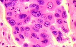
SMALL (OAT) CELL CARCINOMA
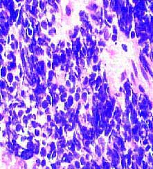
Small (Oat) cell carcinoma accounts for about 20-30% of all lung cancers, these cells secrete a large amount of polypeptide hormones, thought to be as a result of their development from cells of the APUD system. This produces extra-pulmonary manifestations such as SIADH and ectopic adrenocorticotrophin syndrome. This form of lung cancer is responsive to chemotherapy. Under the microscope they form sheets of darkly staining cells with prominent nuclei and little cytoplasm,8 their secretory activity can be seen as the presence of neurosecretory granules in the cytoplasm seen by electron microscopy. This form is very strongly linked to smoking as a causative factor.
SQUAMOUS CELL CARCINOMA
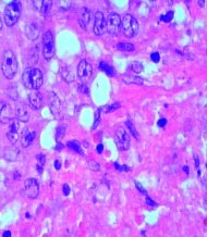
Squamous cell carcinoma (epidermoid carcinoma) accounts for about 30 or 40% of primary lung tumours. They grow most commonly in the central areas in or around major bronchi. They grow in a stratified or pseudoductal arrangement, the cells have an epithelial pearl formation with individual cell keratinization. These tumours deposit keratin, and as they grow develop a necrotic, keratinous mass which appears cheesy on dissection.8 Widespread Metastases occur relatively late. Some results can be achieved if slow growing and treated with radiotherapy.
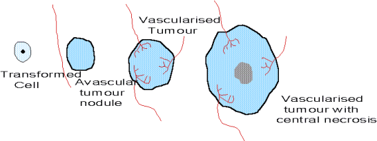 As a tumour grows it secretes substances called tumour angiogenesis factors (TAF) which cause
blood vessels to grow into the mass of tumour cells. This allows a tumour to grow more rapidly and increase in size.
If the tumour grows too large for its blood supply then the central areas can become deprived of oxygen and nutrients
and will undergo necrosis, they die.
This is the process by which squamous cell carcinomas frequently develop into necrotic masses.
Some work into the treatment of various cancers has looked at the possibility of suppressing or counteracting
the effect of TAF in the hope that restricting the flow of blood to a tumour will restrict growth and spread.9
As a tumour grows it secretes substances called tumour angiogenesis factors (TAF) which cause
blood vessels to grow into the mass of tumour cells. This allows a tumour to grow more rapidly and increase in size.
If the tumour grows too large for its blood supply then the central areas can become deprived of oxygen and nutrients
and will undergo necrosis, they die.
This is the process by which squamous cell carcinomas frequently develop into necrotic masses.
Some work into the treatment of various cancers has looked at the possibility of suppressing or counteracting
the effect of TAF in the hope that restricting the flow of blood to a tumour will restrict growth and spread.9
ADENOCARCINOMA
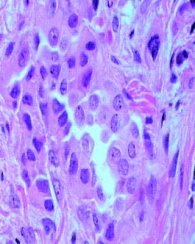
Adenocarcinomas arise peripherally from mucous glands and the cells retain some of the tubular, acinar or papillary differentiation and mucus production.8 They commonly invade pleura and mediastinal lymph nodes and often metastasise to the brain and bones. They bear similarity to secondary tumours and must be distinguished by CT scans and other investigations to check for presence of a primary. Adenocarcinoma commonly arises around scar tissue and is also associated with asbestos exposure. One form of adenocarcinoma is often distinguished from others, bronchiolo-alveolar carcinoma, these characteristically have well differentiated �bland� cells which grow along alveolar ducts. Adenocarcinomas are proportionally less common in non-smokers.
LARGE CELL CARCINOMA
Large cell carcinoma metastasises early and may simply be considered to be those cancers which do not fit into the categories above. Close study by electron microscopy indicates that these types can probably be included with squamous or adenocarcinomas.
MESOTHELIOMAS
Tumours arising from the mesothelium, mesotheliomas, were only well recognised about 25 years ago. The tumour grows over serosal surfaces, and interlobar fissures. The cells are epithelial and spindle types and occur in differing ratios, mitosis is infrequently seen. A common complication of mesothelioma is pleural effusion, particularly recurrent effusion, and it may present as such. Distinction between benign and malignant effusions is difficult. Evidence relating this form of lung cancer to asbestos exposure (particularly crocidolite) has lead to several industrial court cases.10 There appears to be a latent period of between twenty and forty years between exposure and diagnosis.
BENIGN LUNG TUMOURS
Only 3% of lung tumours are benign, most are pulmonary hamartomas or carcinoid tumours and other forms are very rare.
Pulmonary hamartomas are small well-defined non-neoplastic areas of disorganised tissue overgrowth that commonly arise in the periphery of the lung parenchyma. Though not strictly tumours they are included here on the basis of the similarity in appearance on chest X-ray and the subsequent need to differentiate between them and lung cancers. They are very slow growing and generally asymptomatic but may cause obstruction if arising from a major bronchus.
Bronchial carcinoid tumours are very low grade malignant tumours arising in larger bronchi, they grow slowly and eventually cause lobar collapse by blocking the bronchus. The tumour has a typical endocrine packeted appearance, with secretory granules, and resemble intestinal carcinoids. They rarely give rise to the carcinoid syndrome. Adenomas may form in the bronchial mucous gland and can obstruct the bronchioles as they grow.
Benign tumours can arise from any type of tissue in the lung and neurofibromas, lipomas etc may be found in the lung.
Tumour Formation
Tumours arise via a process of carcinogenesis, the most accepted model for this process is the multistage model. Carcinogenic agents affect DNA by several molecular mechanisms, generally they form electrophilic intermediates that bind to DNA causing mispairing or base substitutions during DNA synthesis. It has been demonstrated that in lung cancer large areas �fields� of genetic mutations are found throughout the lung, even when no histological abnormality is seen.11
The first step is when carcinogenic agents mutate DNA to give altered gene expression (initiation). The altered gene expression leads to �promotion,� an initiated cell undergoes clonal expansion to form a preneoplastic lesion. Should this lesion undergo further genetic change (conversion) a malignant tumour develops. The changes that lead to tumour formation occur in the genetic material that is responsible for regulating cell growth and differentiation. Proto-oncogenes and tumour suppressor genes play an important role in the multistage model of carcinogenesis.12 Proto-oncogenes are important to the regulatory mechanisms of growth, cell-cycle control, programmed cell death and terminal differentiation. It has been demonstrated that activation of the ras proto-oncogenes leads to tumour formation,13 angiogenesis14 and metastasis.15 It is thought that K-ras activation is particularly important in the development of adenocarcinomas of the lung.
Tumour suppressor genes code for products that enhance neoplastic activity when mutated. The most commonly altered tumour suppressor gene is the p53 gene found on chromosome 17 ,16,17,18 it codes for a phosphoprotein involved in the control of cell proliferation and loss of function can be caused by single base substitutions.19 It is found to be mutated in 90% of Small Cell lung cancer and 50% of Non Small Cell lung cancer.20
SPREAD
Lung cancers can metastasise to remote sites by blood or lymphatics. Lymphatic spread tends to be more local, often the hilar, mediastinal and peribronchial lymph nodes are affected. Blood borne spread can reach virtually any organ of the body, commonly liver, bones, adrenal glands and brain.
Metastatic spread involves many processes, first the cells must escape the primary site, to do this it must break through the basement membrane to the extracellular matrix (ECM). It is the penetration of the basement layer that distinguishes benign from invasive tumours. Enzymes from the malignant tumour cells are capable of degrading the ECM, these include serine proteases, cathepsins and the matrix metalloproteinases (MMPs).
MMPs have also been suggested as having a part in the control of growth and apoptosis of cells. Work is being done in Leicester on how it may be possible to inhibit these enzymes and prevent metastasis in lung cancer, clinical trials of synthetic inhibitors of MMPs (batimastat, marimastat) are being carried out. These may lead to a new approach in the management of lung cancer.21
As well as metastases, lung cancers often cause local complications of obstruction, ulceration, invasion and systemic effects due to central necrosis and paramalignant disorders. Obstruction of any nearby structure can occur (either by pressure from contained tumour or invasion) and often include SVC obstruction, vocal cord paralysis due to damage to recurrent laryngeal nerve, dyspnoea due to lobar collapse, and dysphagia. Occasionally central necrosis occurs in the middle of a tumour (particularly in squamous carcinoma) when it outgrows its blood supply, the release of pyrogens causes fever in the absence of infection. Paraneoplastic disorders are found in lung cancers with ectopic ACTH production, SIADH, cachexia, clubbing and anaemia found in some cases.